ITN and DAIC Editor Dave Fornell takes a tour of some of the most innovative new technologies being displayed on the expo floor at the Radiological Society of North America (RSNA) 2016 meeting.For key take away trends at RSNA, watch the video "Key Trends, New Technology at RSNA 2016."
236 ViewsRecent VideosView all 642 items

Dr.Frank A. Vicini,MD, FACR, FASTRO, FABS, who is a radiation oncologist withGenesisCare, discussed his findings fromaphase III trial presented atASTRO22that evaluated new and improved techniques to treat patients with breast cancer. Here he shares some of his findings withImaging Technology News (ITN).
Related Conference Coverage:
Photo Gallery of Technologies Showcased at ASTRO 2022
ASTRO 2022 Shines Spotlight on “Cancer Breakthroughs” with AAPM, ASCO Research
“Cancer Breakthroughs” Session at ASTRO2022 Unveils Key Findings from ASCO, AAPM
Looking Ahead to ASTRO: Keynotes, Awards and Scientific Session Updates

Digital solutions like AI and the cloud are driving innovation and transforming the way radiologists and imaging centers approach their work.ITNrecently spoke with Alexandre Salvador, vice president and global head of digital business solutions for Bayer's radiology business, aboutCalantic Digital Solutions, Bayer's newcloud-based platformproviding AI applications integrated into the clinical workflow of the radiology department.

John C. Breneman, M.D., medical director of theCincinnati Children's/UC Health Proton Therapy Center, and the principal investigator of theFAST-01 trial,explains FLASH therapy and details on the trial. This study is testing the use of a single FLASH proton therapy session, rather than weeks of fractionated doses.
FLASH and hypo fractionated therapy have been among the hottest topics in radiation oncology. The premise of FLASH is to deliver extremely high doses of radiation to a tumor in one, short dose. Lab testing has shown this actually has a healthy tissue sparing capability and may help in reducing collateral damage.
If this and other trials show benefit and improved outcomes from FLASH, it is possible this may become the primary treatment method for many cancers in the years to come. Reducing therapy to one treatment session also would open up much more time for proton centers so many more patients could be treated. It also would be a significant time and cost savings for patients and their families, who would not be required to stay at nearby hotels for extended stays during their course of treatment.
Cincinnati Children's/UC Health Proton Therapy Center announced the completion of enrollment in FAST-01 (FeAsibility Study of FLASH Radiotherapy for the Treatment of Symptomatic Bone Metastases) in October. This is the first human clinical trial of FLASH therapy, which centered on patients with metastases in arms and legs to avoid irradiating critical structures. If this trials shows benefits and low toxicity, followup studies will attempt more complex treatments in other parts of the body.
Related ASTRO 2021 Radiation Oncology Content:
7 Trends in Radiation Therapy at ASTRO 2021
Radiation Oncology Research Featured at ASTRO 2021
Photo Gallery of Technologies at ASTRO 2021
VIDEO: Sedating Children With Movies Rather Than Drugs for Radiation Therapy— Interview with Jeffrey T. Chapman
VIDEO: 4 Radiation Oncology Technologies to Watch— Interview with Anthony Zietman, M.D.
VIDEO: Advances in Radiopharmaceutical Therapy— Interview with Ana Kiess, M.D., Ph.D.
VIDEO: MRI-Linac and PSMA PET Imaging Technologies Aids Therapy at GenesisCare— Interview with Walter Curran, Jr. M.D.,
VIDEO: Elekta Harmony Radiotherapy System Walk-around
VIDEO Example of the Varian Noona Bidirection Oncology Patient Interface Software
视频:例子啊f Cherenkov Radiation Imaging During Radiation Therapy

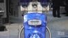
Arun Nagdev, M.D., director of emergency ultrasound at theAlameda Health System, clinical associate professor, University of California San Francisco (UCSF) School of Medicine, and incoming president for theAmerican College of Emergency Physicians(ACEP) ultrasound section, explains the rise inpoint-of-care ultrasound(POCUS) since the start of theCOVID-19pandemic. Nagdev has used cart-based and several hand-held systems to the emergency department to triage patients, help sort out COVID and non-COVID patients and identify patients that need more detailed radiology imaging or with issues that need immediate attention, such as pulmonary embolism, heart attacks and aortic aneurisms.
Nagdev also serves as senior director of clinical education for start-up POCUS companyExo, which is developing a new chip-based, handheld ultrasound system with a high definition transducer.
Watch the relatedVIDEO: How to Image COVID-19 and Radiological Presentations of the Virus— Interview with Margarita Revzin, M.D.
Sponsored VideosView all 171 items
Digital solutions like AI and the cloud are driving innovation and transforming the way radiologists and imaging centers approach their work.ITNrecently spoke with Alexandre Salvador, vice president and global head of digital business solutions for Bayer's radiology business, aboutCalantic Digital Solutions, Bayer's newcloud-based platformproviding AI applications integrated into the clinical workflow of the radiology department.
Software automation can help improve many processes, including verifying eligibility for patient exams, navigating the patient responsibility landscape, and meeting the upcomingCDSMmandate for Appropriate Use Criteria.ITNrecently spoke with with Kevin Borden, Vice President of Product, HCIT, forKonica Minolta Healthcare Americasabout how Konica Minolta is leveraging automation to enhance productivity and efficiency in these areas.
Related content:
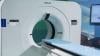
AtRSNA 2021, Philips highlighted the launch of two new innovativeCT systems– the multi-energySpectral CT 7500and theCT 5100 Incisivewith embeddedAIcapabilities.ITNspoke with Wendy Winkle Lawless, CT Business Market Leader - North America, Philips, to learn more about these new systems.
With the use ofcontrast agentsandradiotracers在上升,GE Healthcarehas seen increases in demand across their pharmaceutical diagnostics business. AtRSNA 2021总经理,马可Campione、制药Diagnostics Americas, GE Healthcare, shared how recent investments are helping to meet this increased demand.
Technology ReportsView all 12 items
This is an overview of trends and technologies in radiologyartificial intelligence (AI)applications in 2021. Views were shared by 11 radiologists using AI and industry leaders, which include:
•Randy Hicks, M.D.,MBA, radiologist and CEO of Reginal Medical Imaging (RMI), and an iCAD Profound AI user.
•Prof. Dr. Thomas Frauenfelder, University of Zurich, Institute for Diagnostic and Interventional Radiology, and Riverain AI user.
•Amy Patel, M.D., medical director of Liberty Hospital Women’s Imaging, assistant professor of radiology at UMKC, and user of Kios AI for breast ultrasound.
•Sham Sokka, Ph.D., vice president and head of innovation, precision diagnosis, Philips Healthcare.
•Ivo Dreisser, Siemens Healthineers, global marketing manager for the AI Rad Companion.
•Bill Lacey,vice president of medical informatics, Fujifilm Medical Systems USA.
•Karley Yoder,vice president and general manager, artificial intelligence, GE Healthcare.
•Georges Espada,head of Agfa Healthcare digital and computed radiography business unit.
•Pooja Rao, head of research and development and co-founder of Qure.ai.
•Jill Hamman,world-wide marketing manager at Carestream Health.
•Sebastian Nickel, Siemens Healthineers, global product manager for the AI Pathway Companion.
There has been a change in attitudes about AI on the expo floor at theRadiological Society of North America(RSNA)在过去的两年里。人工智能对话were originally 101 level and discussed how AI technology could be trained to sort photos of dogs and cats. However, in 2020, with numerous FDA approvals for various AI applications, the conversations at RSNA, and industry wide, have shifted to that of accepting the validity of AI. Radiologists now want to discuss how a specific AI algorithm is going to help them save time, make more accurate diagnoses and make them more efficient.
With a higher level of maturity in AI and the technology seeing wider adoption, radiologists using it say AI gives them additional confidence in their diagnoses, and can even help readers who may not be deep experts in the exam type they are being asked to read.
With a myriad of new AI apps gaining regulatory approval from scores of imaging vendors, the biggest challenge for getting this technology into hospitals is an easy to integrate format. This has led to several vendors creating AI app stores. These allow AI apps to integrate easily into radiology workflows because the apps are already integrated as third-party software into a larger radiology vendors' IT platform.
There are now hundreds of AI applications that do a wide variety of analysis, from data analytics, image reconstruction, disease and anatomy identification, automating measurements and advanced visualization. The AI applications can be divided into 2 basic types — AI to improve workflow, and AI for clinical decision support, such as diagnostic aids.
On the workflow side, several vendors are leveraging AI to pull together all of a patients' information, prior exams and reports in one location and to digest the information so it is easier for the radiologist to consume. Often the AI pulls only data and priors that relate to a specific question being asked, based on the imaging protocol used for the exam. One example of this is the Siemens Healthineers AI Clinical Pathway and Siemens AI integrations with PACS to automate measurements and advanced visualization.
AI is also helping simplify complex tasks and help reduce the reading time on involved exams. One example of this is in 3-D breast tomosythesis with hundreds of images, which is rapidly replacing 2-D mammography, which only produces 4 images. Another example is automated image reconstruction algorithms to significantly reduce manual work. AI also is now being integrated directly into several vendors' imaging systems to speed workflow and improve image quality.
Vendors say AI is here to stay. They explain the future of AI will be automation to help improve image quality, simplify manual processes, improved diagnostic quality, new ways to analyze data, and workflow aids that operate in the background as part of a growing number of software solutions.
Several vendors at RSNA 2020 noted that AI's biggest impact in the coming years will be its ability to augment and speed the workflow for the small number of radiologists compared to the quickly growing elder patient populations worldwide. There also are applications in rural and developing countries were there are very low numbers of physicians or specialists.
Related AI in Medical Imaging Content:
AI Outperforms Humans in Creating Cancer Treatments, But Do Doctors Trust It?
VIDEO: Artificial Intelligence For MRI Helps Overcome Backlog of Exams Due to COVID
How AI is Helping the Fight Against Breast Cancer
VIDEO: Use of Artificial Intelligence in Nuclear Imaging
3 High-impact AI Market Trends in Radiology at RSNA 2019
Photo Gallery of New Imaging Technologies at RSNA 2019
VIDEO: Editors Choice of the Most Innovative New Radiology Technology at RSNA 2019
Study Reveals New Comprehensive AI Chest X-ray Solution Improves Radiologist Accuracy
VIDEO: Real-world Use of AI to Detect Hemorrhagic Stroke
The Radiology AI Evolution at RSNA 2019
Eliminating Bias from Healthcare AI Critical to Improve Health Equity
VIDEO: FDA Cleared Artificial Intelligence for Immediate Results of Head CT Scans
Building the Future of AI Through Data
Integrating Artificial Intelligence in Treatment Planning
Selecting an AI Marketplace for Radiology: Key Considerations for Healthcare Providers
Artificial Intelligence Improves Accuracy of Breast Ultrasound Diagnoses
Artificial Intelligence Greatly Speeds Radiation Therapy Treatment Planning
研讨会:建造这座桥——成像AI是Delivering Clinical Value Across the Care Continuum
AI in Medical Imaging Market to Reach $1.5B by 2024
VIDEO: AI-Assisted Automatic Ejection Fraction for Point-of-Care Ultrasound
5 Trends in Enterprise Imaging and PACS Systems
VIDEO: Artificial Intelligence to Automate CT Calcium Scoring and Radiomics
Scale AI in Imaging Now for the Post-COVID Era
VIDEO: Integrating Artificial Intelligence Into Radiologists Workflow
Northwestern Medicine Introduces Artificial Intelligence to Improve Ultrasound Imaging

In Artificial Intelligence at RSNA 2019, ITN Contributing Editor Greg Freiherr offers an overview of artificial intelligence (AI) advances at the Radiological Society of North America (RSNA) 2019 annual meeting.

InEnterprise Imaging at RSNA 2019,ITNContributing Editor Greg Freiherr offers an overview of enterprise imaging advances at the Radiological Society of North America (RSNA) 2019 annual meeting.

InArtificial Intelligence 2018: What Radiologists Need to Know About AI,ITNContributing EditorGreg Freiherroffers an overview ofartificial intelligence (AI)advances at theRadiological Society of North America (RSNA)2018 annual meeting.
Related Artificial Intelligence Content
Technology Report: Artificial Intelligence 2017
VIDEO: RSNA Post-game Report on Artificial Intelligence
VIDEO: AI in Tumor Diagnostics, Treatment and Follow-up
VIDEO: Artificial Intelligence May Help Reduce Gadolinium Dose in MRI
Radiation OncologyView all 137 items
Dr.Frank A. Vicini,MD, FACR, FASTRO, FABS, who is a radiation oncologist withGenesisCare, discussed his findings fromaphase III trial presented atASTRO22that evaluated new and improved techniques to treat patients with breast cancer. Here he shares some of his findings withImaging Technology News (ITN).
Related Conference Coverage:
Photo Gallery of Technologies Showcased at ASTRO 2022
ASTRO 2022 Shines Spotlight on “Cancer Breakthroughs” with AAPM, ASCO Research
“Cancer Breakthroughs” Session at ASTRO2022 Unveils Key Findings from ASCO, AAPM
Looking Ahead to ASTRO: Keynotes, Awards and Scientific Session Updates
John C. Breneman, M.D., medical director of theCincinnati Children's/UC Health Proton Therapy Center, and the principal investigator of theFAST-01 trial,explains FLASH therapy and details on the trial. This study is testing the use of a single FLASH proton therapy session, rather than weeks of fractionated doses.
FLASH and hypo fractionated therapy have been among the hottest topics in radiation oncology. The premise of FLASH is to deliver extremely high doses of radiation to a tumor in one, short dose. Lab testing has shown this actually has a healthy tissue sparing capability and may help in reducing collateral damage.
If this and other trials show benefit and improved outcomes from FLASH, it is possible this may become the primary treatment method for many cancers in the years to come. Reducing therapy to one treatment session also would open up much more time for proton centers so many more patients could be treated. It also would be a significant time and cost savings for patients and their families, who would not be required to stay at nearby hotels for extended stays during their course of treatment.
Cincinnati Children's/UC Health Proton Therapy Center announced the completion of enrollment in FAST-01 (FeAsibility Study of FLASH Radiotherapy for the Treatment of Symptomatic Bone Metastases) in October. This is the first human clinical trial of FLASH therapy, which centered on patients with metastases in arms and legs to avoid irradiating critical structures. If this trials shows benefits and low toxicity, followup studies will attempt more complex treatments in other parts of the body.
Related ASTRO 2021 Radiation Oncology Content:
7 Trends in Radiation Therapy at ASTRO 2021
Radiation Oncology Research Featured at ASTRO 2021
Photo Gallery of Technologies at ASTRO 2021
VIDEO: Sedating Children With Movies Rather Than Drugs for Radiation Therapy— Interview with Jeffrey T. Chapman
VIDEO: 4 Radiation Oncology Technologies to Watch— Interview with Anthony Zietman, M.D.
VIDEO: Advances in Radiopharmaceutical Therapy— Interview with Ana Kiess, M.D., Ph.D.
VIDEO: MRI-Linac and PSMA PET Imaging Technologies Aids Therapy at GenesisCare— Interview with Walter Curran, Jr. M.D.,
VIDEO: Elekta Harmony Radiotherapy System Walk-around
VIDEO Example of the Varian Noona Bidirection Oncology Patient Interface Software
视频:例子啊f Cherenkov Radiation Imaging During Radiation Therapy
Elekta’s latest linear accelerator,Harmony, is designed to provide a productive and versatile radiotherapy solution for both mature and developing markets.ITNrecently spoke with Chris Gilpin, Global Product Marketing Manager, and Emily Basset, Global Clinical Marketing Manager, to learn more about the treatment system.
Related content:
核医学和辐射其他的使用日益增加apy for Diagnosis and Treatment
7 Trends in Radiation Therapy at ASTRO 2021
VIDEO: Elekta Harmony Radiotherapy System Walk-around
Photo Gallery of Technologies at ASTRO 2021
Magdalena Bazalova-Carter, Ph.D., assistant professor, University of Victoria University, discusses the current state of ultra-high dose FLASHradiation therapyat the 2021American Society of Radiation Oncology (ASTRO)annual meeting. Flash therapy is said to be a key technology to keep an eye on in the next few years. If it proves viable in human patients, it promises to greatly shorten treatment times, and reduce fractions to between 1-3 sessions.
The idea is that a super-high dose of radiation is delivered in one large, very fast dose. It appears that despite the high dose of radiation, there is a tissue sparing biology mechanism that is not yet fully understood, where health tissue is preserved and there is less collateral damage than the standard series of lower dose fractions over days or weeks.
Flash therapy is being tested in electron beam therapy systems to treat superficial cancers, which are much easier to adopt to flash than deeper tissue tumors. Proton may be able to produce the higher energies needed for deeper tumor treatments, but current photon beam systems are limited because to deliver the high doses needed may cause enough heat to melt the X-ray beam source.
7 Trends in Radiation Therapy at ASTRO 2021
Photo Gallery of Technologies at ASTRO 2021
Radiology ImagingView all 386 items
Dr.Frank A. Vicini,MD, FACR, FASTRO, FABS, who is a radiation oncologist withGenesisCare, discussed his findings fromaphase III trial presented atASTRO22that evaluated new and improved techniques to treat patients with breast cancer. Here he shares some of his findings withImaging Technology News (ITN).
Related Conference Coverage:
Photo Gallery of Technologies Showcased at ASTRO 2022
ASTRO 2022 Shines Spotlight on “Cancer Breakthroughs” with AAPM, ASCO Research
“Cancer Breakthroughs” Session at ASTRO2022 Unveils Key Findings from ASCO, AAPM
Looking Ahead to ASTRO: Keynotes, Awards and Scientific Session Updates
Arun Nagdev, M.D., director of emergency ultrasound at theAlameda Health System, clinical associate professor, University of California San Francisco (UCSF) School of Medicine, and incoming president for theAmerican College of Emergency Physicians(ACEP) ultrasound section, explains the rise inpoint-of-care ultrasound(POCUS) since the start of theCOVID-19pandemic. Nagdev has used cart-based and several hand-held systems to the emergency department to triage patients, help sort out COVID and non-COVID patients and identify patients that need more detailed radiology imaging or with issues that need immediate attention, such as pulmonary embolism, heart attacks and aortic aneurisms.
Nagdev also serves as senior director of clinical education for start-up POCUS companyExo, which is developing a new chip-based, handheld ultrasound system with a high definition transducer.
Watch the relatedVIDEO: How to Image COVID-19 and Radiological Presentations of the Virus— Interview with Margarita Revzin, M.D.
AtRSNA 2021, Philips highlighted the launch of two new innovativeCT systems– the multi-energySpectral CT 7500and theCT 5100 Incisivewith embeddedAIcapabilities.ITNspoke with Wendy Winkle Lawless, CT Business Market Leader - North America, Philips, to learn more about these new systems.

AtRSNA 2021, Konica Minolta introduced themKDR XpressMobile X-ray system and theAero DR Carbon flat panel detector. Also on display wasnVoq’s cloud-based speech recognition and automation solution and new features for theExa platformthat automate common clinical and administrative tasks.
Molecular ImagingView all 33 items
AtRSNA 2021, Philips highlighted the launch of two new innovativeCT systems– the multi-energySpectral CT 7500and theCT 5100 Incisivewith embeddedAIcapabilities.ITNspoke with Wendy Winkle Lawless, CT Business Market Leader - North America, Philips, to learn more about these new systems.
Ana Kiess, M.D., Ph.D., assistant professor ofradiation oncologyand molecular radiation sciences, Johns Hopkins University, explains the current state of patient-centered radiopharmaceutical therapy at theAmerican Society of Radiation Oncology (ASTRO)2021年的会议。
She discusses development and use over the past decade of Radium-223 dichloride and Lutetium-177 dotatate. Kiess also expects there will be targeted injectable radiopharmaceuticals for nearly all solid tumor types in the next decade. She said her center is currently investigating the use of radiopharma agents to treat oligometastatic metastatic cancers.
Photo Gallery of Technologies at ASTRO 2021

Walter Curran, Jr. M.D., FACR, FASCO, GenesisCare global chief medical officer, discusses three technologies that are helping advance radiation oncology care during theAmerican Society of Radiation Oncology (ASTRO)2021年的会议。These technology advances include:
• Prostate-specific membrane antigen (PSMA) positron emission tomography (PET) imaging for prostate cancer.
• MRI-linear accelerator (Linac) systems that allow real-time imaging during radiation therapy.
• Remote treatment planning to help radiation treatment centers that are in rural areas.
Prostate cancer is one of the most common cancers in men. The new PSMA PET imaging is expected to significantly improve how prostate cancer is detected and treated. The FDA approved the drug for PET nuclear imaging of PSMA-positive lesions in men with prostate cancer. 68Ga-PSMA-11 is a radioactive imaging agent that binds to prostate cancer cells to help localize prostate cancer cells.
Photo Gallery of Technologies at ASTRO 2021

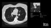
Yael Eshet, M.D., MSc, a diagnostic radiology specialist atSheba Medical Centerin Israel, was the lead author on a recent study that showedCOVID-19 (SARS-CoV-2)vaccine adenopathy can persist more than 6 weeks. This swelling of lymph nodes is similar to what is seen cancer and infections and the new findings show it can last longer than 7-10 weeks. The current recommended time people should delay medical imaging is 6 weeks after receiving a COVID vaccine to avoid a misdiagnosis,[2] but this new study shows there is increased inflammation shown on PET-CT imaging for much longer.
These were the findings in theRadiologypublished study“患病率增加FDG PET / CT腋窝淋巴结Node Uptake Beyond 6 Weeks after mRNA COVID-19 Vaccination."[1]
Researchers using fluorodeoxyglucose (FDG)-positron emission tomography (PET)have found increased FDG uptake in the lymph nodes of patients 7-10 weeks past their second mRNA-based Pfizer-BioNTech COVID-19 vaccination. This new information indicates a persistent immune response that could be mistaken on imaging exams for serious conditions like lymphoma over a much longer period of time.
Recent recommendations for post-vaccine lymphadenopathy advise scheduling routine imaging, such as screening mammography, before, or at least 6 weeks after, the final vaccination dose to eliminate false positive results. However, this new research showed that avid axillary lymph node uptake was present beyond 6 weeks after the second vaccination in more than 29% of the patients in the study cohort.
The authors stated “This study shows that avid axillary lymph node uptake on FDG PET/CT can be detected in more than a quarter of our patient population even beyond 6 weeks after the second dose of the mRNA-based COVID-19 vaccination. Compared to a previous study showing normalization of FDG uptake within 40 days of receiving an inactivated H1N1 influenza vaccine, we found uptake persistence even at 70 days. Physicians should be aware of this potential pitfall.”
Some images in this video are from anotherRadiologystudy, which showed PET tracer uptake at the COVID vaccine injection site and other examples of axillary adenopathy.[3]
Related COVID Vaccine Axillary Adenapathy Content:
COVID-19 Vaccine Can Cause False Positive Cancer Diagnosis
Help Spread Awareness of Potential COVID-19 Vaccine Imaging Side-effects
VIDEO: COVID Vaccine May Cause Enlarged Lymph Nodes on Mammograms— Interview with Constance "Connie" Lehman, M.D.
COVID-19 Vaccination Axillary Adenopathy Detected During Breast Imaging
PHOTO GALLERY: How COVID-19 Appears on Medical Imaging
CMS Now Requires COVID-19 Vaccinations for Healthcare Workers by January 4
Find more radiology related COVID content
References:
2022年世界杯半区对阵View all 515 items
Dr.Frank A. Vicini,MD, FACR, FASTRO, FABS, who is a radiation oncologist withGenesisCare, discussed his findings fromaphase III trial presented atASTRO22that evaluated new and improved techniques to treat patients with breast cancer. Here he shares some of his findings withImaging Technology News (ITN).
Related Conference Coverage:
Photo Gallery of Technologies Showcased at ASTRO 2022
ASTRO 2022 Shines Spotlight on “Cancer Breakthroughs” with AAPM, ASCO Research
“Cancer Breakthroughs” Session at ASTRO2022 Unveils Key Findings from ASCO, AAPM
Looking Ahead to ASTRO: Keynotes, Awards and Scientific Session Updates
John C. Breneman, M.D., medical director of theCincinnati Children's/UC Health Proton Therapy Center, and the principal investigator of theFAST-01 trial,explains FLASH therapy and details on the trial. This study is testing the use of a single FLASH proton therapy session, rather than weeks of fractionated doses.
FLASH and hypo fractionated therapy have been among the hottest topics in radiation oncology. The premise of FLASH is to deliver extremely high doses of radiation to a tumor in one, short dose. Lab testing has shown this actually has a healthy tissue sparing capability and may help in reducing collateral damage.
If this and other trials show benefit and improved outcomes from FLASH, it is possible this may become the primary treatment method for many cancers in the years to come. Reducing therapy to one treatment session also would open up much more time for proton centers so many more patients could be treated. It also would be a significant time and cost savings for patients and their families, who would not be required to stay at nearby hotels for extended stays during their course of treatment.
Cincinnati Children's/UC Health Proton Therapy Center announced the completion of enrollment in FAST-01 (FeAsibility Study of FLASH Radiotherapy for the Treatment of Symptomatic Bone Metastases) in October. This is the first human clinical trial of FLASH therapy, which centered on patients with metastases in arms and legs to avoid irradiating critical structures. If this trials shows benefits and low toxicity, followup studies will attempt more complex treatments in other parts of the body.
Related ASTRO 2021 Radiation Oncology Content:
7 Trends in Radiation Therapy at ASTRO 2021
Radiation Oncology Research Featured at ASTRO 2021
Photo Gallery of Technologies at ASTRO 2021
VIDEO: Sedating Children With Movies Rather Than Drugs for Radiation Therapy— Interview with Jeffrey T. Chapman
VIDEO: 4 Radiation Oncology Technologies to Watch— Interview with Anthony Zietman, M.D.
VIDEO: Advances in Radiopharmaceutical Therapy— Interview with Ana Kiess, M.D., Ph.D.
VIDEO: MRI-Linac and PSMA PET Imaging Technologies Aids Therapy at GenesisCare— Interview with Walter Curran, Jr. M.D.,
VIDEO: Elekta Harmony Radiotherapy System Walk-around
VIDEO Example of the Varian Noona Bidirection Oncology Patient Interface Software
视频:例子啊f Cherenkov Radiation Imaging During Radiation Therapy
Software automation can help improve many processes, including verifying eligibility for patient exams, navigating the patient responsibility landscape, and meeting the upcomingCDSMmandate for Appropriate Use Criteria.ITNrecently spoke with with Kevin Borden, Vice President of Product, HCIT, forKonica Minolta Healthcare Americasabout how Konica Minolta is leveraging automation to enhance productivity and efficiency in these areas.
Related content:
AtRSNA 2021, Philips highlighted the launch of two new innovativeCT systems– the multi-energySpectral CT 7500and theCT 5100 Incisivewith embeddedAIcapabilities.ITNspoke with Wendy Winkle Lawless, CT Business Market Leader - North America, Philips, to learn more about these new systems.
沙特vs阿联酋比分预测View all 281 items
Digital solutions like AI and the cloud are driving innovation and transforming the way radiologists and imaging centers approach their work.ITNrecently spoke with Alexandre Salvador, vice president and global head of digital business solutions for Bayer's radiology business, aboutCalantic Digital Solutions, Bayer's newcloud-based platformproviding AI applications integrated into the clinical workflow of the radiology department.
Software automation can help improve many processes, including verifying eligibility for patient exams, navigating the patient responsibility landscape, and meeting the upcomingCDSMmandate for Appropriate Use Criteria.ITNrecently spoke with with Kevin Borden, Vice President of Product, HCIT, forKonica Minolta Healthcare Americasabout how Konica Minolta is leveraging automation to enhance productivity and efficiency in these areas.
Related content:
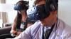
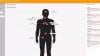
Douglas E. Holt, M.D., a radiation oncologist atEastern Idaho Regional Medical Center,explains the use of 3-D virtual reality volumetric imaging review to help improve cancer patients’ understanding of their disease and treatment. Pictures are worth a thousand words, and moving pictures inside a patient's body even more. Holt said using virtual reality to go through the patient's anatomy in 3D and to show them what is wrong and how it will be treated offers a new level of understanding that is not possible using a discussion or a couple still images from their medical imaging.
Holt presented this study as a late-breaker at the 2021American Society of Radiation Oncology (ASTRO)annual meeting.
Find more ASTRO videos and news
7 Trends in Radiation Therapy at ASTRO 2021
Photo Gallery of Technologies at ASTRO 2021

Rik Primo, principle atPrimo Medical Imaging Informatics Consultantsand former health IT developer with Siemens, Philips and Agfa, explains the difference cloud-native versus cloud-enabled PACS and放射学企业成像系统. He spoke withITNduring RSNA 2021.theRadiological Society of North America (RSNA)2021 annual meeting.
Women's HealthView all 79 items
Dr.Frank A. Vicini,MD, FACR, FASTRO, FABS, who is a radiation oncologist withGenesisCare, discussed his findings fromaphase III trial presented atASTRO22that evaluated new and improved techniques to treat patients with breast cancer. Here he shares some of his findings withImaging Technology News (ITN).
Related Conference Coverage:
Photo Gallery of Technologies Showcased at ASTRO 2022
ASTRO 2022 Shines Spotlight on “Cancer Breakthroughs” with AAPM, ASCO Research
“Cancer Breakthroughs” Session at ASTRO2022 Unveils Key Findings from ASCO, AAPM
Looking Ahead to ASTRO: Keynotes, Awards and Scientific Session Updates
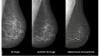
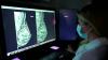
Stamatia Destounis, M.D., FACR, chief of the American College of Radiology (ACR) Breast Commission, managing partner, Elizabeth Wende Breast Care, Rochester, N.Y., explains some of the key trends in2022世界杯篮球预选赛赛程at the 2021Radiological Society of North America (RSNA)meeting.
She discusses the trends of 3D mammography seeing rapid growth, adoption of synthetic 2D breast images from the tomosynthesis datasets, contrast-enhanced mammography, and breast MRI to help women with dense breast tissue. Destounis also discusses the use of artificial intelligence (AI) to help radiologists with finding what they needs with larger datasets in 3D mammography, and to help act as a second set of eyes.
Early in 2021, with the roll out of the COVID vaccines, one of the biggest headlines in radiology was that the vaccine can show false positives for cancer because it may cause inflammation of lymph nodes. Destounis explains this issue and how women's health centers have largely overcome this by asking patients about their vaccination status and planning imaging around the vaccination dates.
Related Breast Imaging Content:
COVID-19 Vaccine Can Cause False Positive Cancer Diagnosis
Help Spread Awareness of Potential COVID-19 Vaccine Imaging Side-effects
VIDEO: COVID Vaccine May Cause Enlarged Lymph Nodes on Mammograms— Interview with Constance "Connie" Lehman, M.D.
COVID-19 Vaccination Axillary Adenopathy Detected During Breast Imaging
VIDEO: COVID Vaccine Adenopathy Can Last Up to 10 Weeks— Interview with Yael Eshet, M.D.
VIDEO: Artificial Intelligence Trends in Medical Imaging— Interview with Signify Research
Technology Report: Artificial Intelligence in Radiology 2021
Yael Eshet, M.D., MSc, a diagnostic radiology specialist atSheba Medical Centerin Israel, was the lead author on a recent study that showedCOVID-19 (SARS-CoV-2)vaccine adenopathy can persist more than 6 weeks. This swelling of lymph nodes is similar to what is seen cancer and infections and the new findings show it can last longer than 7-10 weeks. The current recommended time people should delay medical imaging is 6 weeks after receiving a COVID vaccine to avoid a misdiagnosis,[2] but this new study shows there is increased inflammation shown on PET-CT imaging for much longer.
These were the findings in theRadiologypublished study“患病率增加FDG PET / CT腋窝淋巴结Node Uptake Beyond 6 Weeks after mRNA COVID-19 Vaccination."[1]
Researchers using fluorodeoxyglucose (FDG)-positron emission tomography (PET)have found increased FDG uptake in the lymph nodes of patients 7-10 weeks past their second mRNA-based Pfizer-BioNTech COVID-19 vaccination. This new information indicates a persistent immune response that could be mistaken on imaging exams for serious conditions like lymphoma over a much longer period of time.
Recent recommendations for post-vaccine lymphadenopathy advise scheduling routine imaging, such as screening mammography, before, or at least 6 weeks after, the final vaccination dose to eliminate false positive results. However, this new research showed that avid axillary lymph node uptake was present beyond 6 weeks after the second vaccination in more than 29% of the patients in the study cohort.
The authors stated “This study shows that avid axillary lymph node uptake on FDG PET/CT can be detected in more than a quarter of our patient population even beyond 6 weeks after the second dose of the mRNA-based COVID-19 vaccination. Compared to a previous study showing normalization of FDG uptake within 40 days of receiving an inactivated H1N1 influenza vaccine, we found uptake persistence even at 70 days. Physicians should be aware of this potential pitfall.”
Some images in this video are from anotherRadiologystudy, which showed PET tracer uptake at the COVID vaccine injection site and other examples of axillary adenopathy.[3]
Related COVID Vaccine Axillary Adenapathy Content:
COVID-19 Vaccine Can Cause False Positive Cancer Diagnosis
Help Spread Awareness of Potential COVID-19 Vaccine Imaging Side-effects
VIDEO: COVID Vaccine May Cause Enlarged Lymph Nodes on Mammograms— Interview with Constance "Connie" Lehman, M.D.
COVID-19 Vaccination Axillary Adenopathy Detected During Breast Imaging
PHOTO GALLERY: How COVID-19 Appears on Medical Imaging
CMS Now Requires COVID-19 Vaccinations for Healthcare Workers by January 4
Find more radiology related COVID content
References:
Constance "Connie" Lehman, M.D., Ph.D., chief of breast imaging, co-director of the Avon Comprehensive Breast Evaluation Center at the Massachusetts General Hospital, and professor of radiology at Harvard Medical School, explains issues and suggested guidelines for women who receive the COVID-19 vaccine and need to get a mammogram. In the first three months since the vaccines have been released, there have been numerous case reports of the vaccine causing swollen lymph nodes. This is would usually raise a red flag for breast cancer, but is normal for many women receiving the vaccine as their body's immune system gears up against the virus.
Lehman said cases reports of axillary adenopathy have been identified on breast imaging aftercoronavirus disease (COVID-19)vaccination and are rising. Lehman et al. proposed a pragmatic management approach in arecent article in theAmerican Journal of Roentgenology (AJR).[1]
In the settings of screening mammography, screening MRI and diagnostic imaging work-up of breast symptoms, with no imaging findings beyond unilateral axillary adenopathy ipsilateral to recent (prior six weeks) vaccination, they report the adenopathy as benign with no further imaging indicated if no nodes are palpable six weeks after the last vaccine dose.
For patients with palpable axillary adenopathy in the setting of ipsilateral recent vaccination, clinical follow-up of the axilla is recommended. In all these scenarios, axillary ultrasound is recommended if clinical concern persists six weeks after vaccination.
In patients with recent breast cancer diagnosis in the pre- or peri-treatment setting, prompt recommended imaging is encouraged as well as vaccination (in the thigh or contralateral arm). The recommendations align with the ACR BI-RADS Atlas and aim to: 1) reduce patient anxiety, provider burden, and costs of unnecessary evaluation of enlarged nodes in the setting of recent vaccination, and 2) avoid further delays in vaccinations and breast cancer screening during the pandemic.
Related Medical Imaging of COVID Content:
COVID-19 Vaccination Axillary Adenopathy Detected During Breast Imaging
CMS Now Requires COVID-19 Vaccinations for Healthcare Workers by January 4
PHOTO GALLERY: How COVID-19 Appears on Medical Imaging
VIDEO: Imaging COVID-19 With Point-of-Care Ultrasound (POCUS)— Interview with Mike Stone, M.D.
VIDEO: Use of Teleradiology During the COVID-19 Pandemic— Interview with John Kim, M.D.
Find more radiology related COVID content
Reference: